Lesions causing lameness can be split into two broad categories: infectious and non-infectious, all have slightly different risk factors.
The three most common lesions found on UK dairy farms are, Sole ulcers, White line disease and Digital dermatitis. Sole ulcers and White line disease fall into the non-infectious category
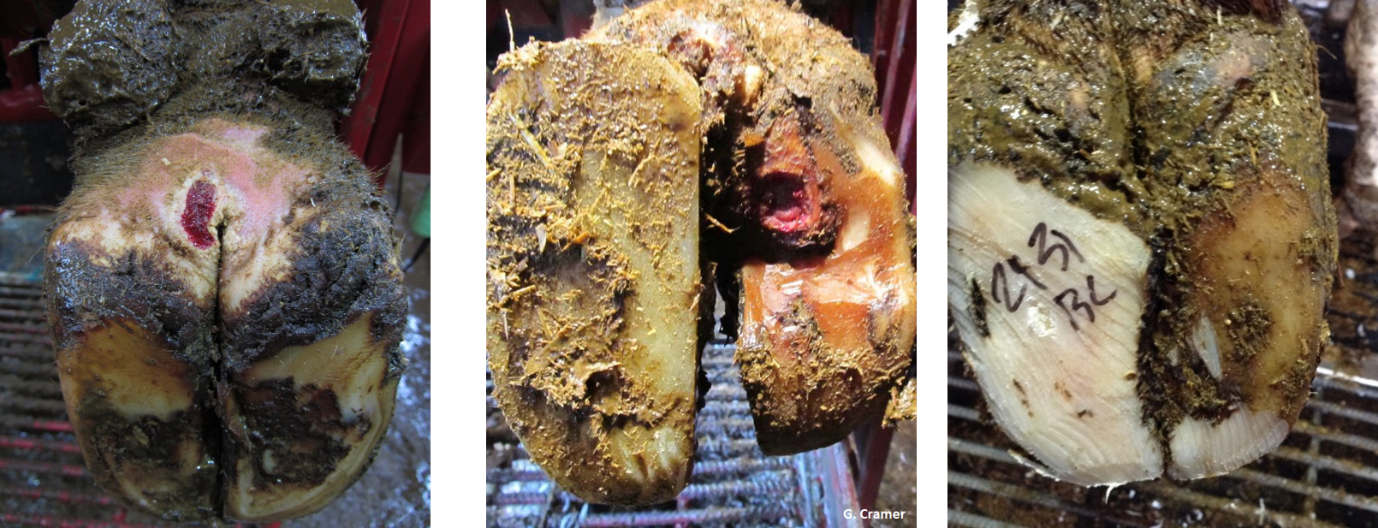
Fig 3: Left to right, Digital Dermatitis, Sole ulcer, White line disease
Sole ulcers
Changes in the foot leading to a sole ulcer typically occurs around calving. This is when laminar attachments in the hoof capsule slacken, allowing P3 to ‘sink’, the flexor tuberosity creates a pinch point between P3 and the ground. Thus compressing the solar corium, disrupting blood flow to keratinocyte cells that produce solar horn. This then produces bruising and inflammation and disrupts sole horn growth. Cows will alter gait to account for the inflammation and claw horn shape is affected, creating a vicious cycle of pinching and bruising. If the pressure is not alleviated then a sole ulcer develops. It takes 2 months for injury at the corium to be seen on the sole, due to sole horn growing roughly 5mm a month. Sole ulcers can be remembered as ‘standing up disease’ due to most of the associated risk factors cause cows to stand for long periods of time.
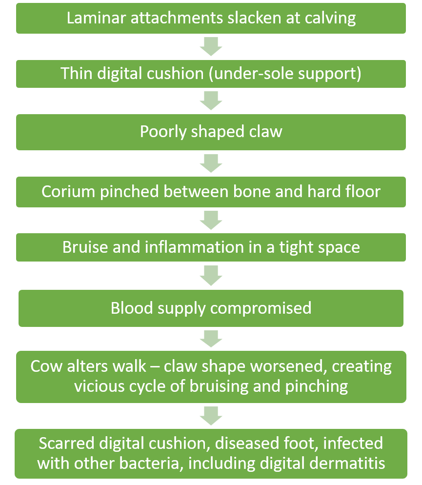
Figure 4: Pathology of Sole ulcers
White line disease
The white line is the junction of wall horn and sole horn and can be seen on the sole when a foot is lifted. It is typically a weak part of the claw. This structure is susceptible to wet conditions, bruising and poor surfaces. Particularly at calving when laminar attachments slacken, the white line can become bruised and horn weakened. Small particles and bacteria can enter the white line. This can cause a painful abscess between the sensitive corium and hard outer wall horn. Risk factors are commonly rough walking surfaces, poor cow flow involving lots of slipping and twisting, bullying or cows pushing each other at feed faces or in collecting yards.
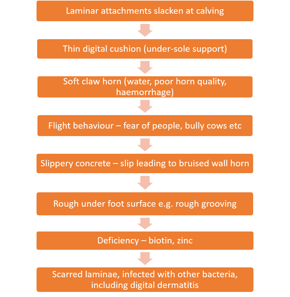
Figure 5: Pathology of White line disease
Treatment of both sole ulcers and white line disease is similar and recent work by Thomas et al (2015) has shown blocks and NSAIDs to me most effective on cure rates.
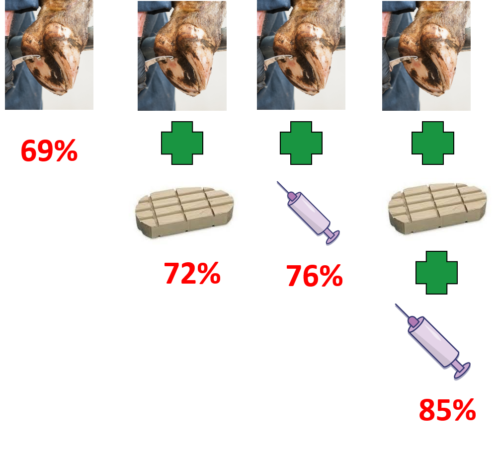
Fig 6: Percentage of cows that were non-lame 5 weeks after treatment. Cure rate was significantly higher in the Trim + block + pain relief group
Full list of foot lesions can be found in ICAR foot lesion atlas.